The Joseph H. and Belle R. Braun Senior Lecturer in Medicine
The brain processes sensory information to extract information that is relevant to guide behavior. We study how such behavioral-relevant information is extracted from odorous scenes. We develop novel behavioral paradigms and combine them with electrophysiology and computational analysis to reveal how olfactory brain circuits extract behavioral-relevant information from complex stimuli.
We are interested in revealing how the brain processes complex odorous stimuli to guide behavior. Many mammalian species rely on the sense of smell for daily basic functions such as finding food, choosing mates, and avoiding predators. In natural settings, odors emitted by these objects will mix with odors emitted from other objects in the environment, prior to reaching the nose. The brain must segment the mixed inputs to be able to detect and identify odors that are behaviorally important. We combine behavioral, electrophysiological, and computational methodologies to reveal the underlying neuronal processes that support the brain’s ability to segment mixtures. We develop behavioral tasks for rodents, that mimic the richness of natural settings, while allowing quantitative analysis, and combine these with dense electrophysiological recordings.

Target-odor detection against background task. Mice are trained to report if a mixture includes a specific target odorant. Trial difficulty is correlated with the glomerular overlap between the target and background odorants.
Similarly to other sensory systems, olfactory brain regions are heavily interconnected via feedforward and feedback connections. While the wiring logic of the feedforward connections has been largely worked-out, that of the feedback connections remains unknown. A major effort in the lab is devoted to understanding the roles played by feedback connections in processing rich and complex odorous stimuli. In addition to electrophysiology during behavior, we also utilize tracing methods and in-vitro preparations to analyze feedback circuit connectivity.

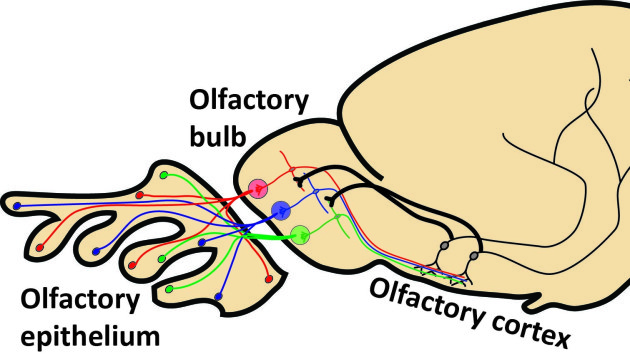
Wiring in the olfactory system. Sensory neurons in the olfactory epithelium express a single olfactory receptor gene (out of ~1300 in the mouse). The projections of sensory neurons expressing the same gene converge in the olfactory bulb to form glomeruli. Mitral and tufted cells, the principal neurons of the olfactory bulb receive inputs from a single glomerulus and therefore represent information from a single receptor type. The olfactory cortex is the first brain station where individual neurons receive converging inputs representing multiple receptor types. The topology of the feedback connections is not yet clear.
Licht, T., Yunerman, M., Maor, I., Lawabny, N., Rokach, R. O., Shiff, I., Mizrahi, A. & Rokni, D (2023) Adaptive olfactory circuitry restores function despite severe olfactory bulb degeneration. Current Biology.
Mammalian olfaction is thought to rely on stereotyped circuitry. Here we describe a mouse model that retains a functional sense of smell despite severe olfactory bulb degeneration. These mice can perform odor-guided tasks and have functional odor representations in piriform cortex, despite extremely aberrant olfactory bulb circuitry.
Lebovich L, Yunerman M, Scaiewicz V, Loewenstein Y, Rokni D (2021) Paradoxical relationship between speed and accuracy in olfactory figure-background segregation. PLoS Comput Biol 17(12): e1009674.
We show that mice respond faster in an olfactory figure-background segmentation task, in trials with more background odors, despite these trials being more difficult for them. This paradoxical relationship between speed and accuracy can be explained by the drift-diffusion model of decision-making if background odors are assumed to act by increasing noise.
Penker D., Licht T., Hofer K.T., and Rokni D. (2020). Mixture Coding and Segmentation in the Anterior Pirifrom Cortex. Front. Syst. Neurosci. 14, 89
Mice can easily detect target odors that are embedded in rich backgrounds. To reveal the neural underpinnings of this ability, we recorded neural responses to single odorants and their mixtures in the piriform cortex. We find that responses to mixtures can be largely predicted from the responses to individual odors using a normalization model. Additionally, we find that mixture components can be decoded from the activity of piriform neurons.
Mathis A.*, Rokni, D.*, Kapoor, V., Bethge, M., and Murthy, V.N. (2016). Reading Out Olfactory Receptors: Feedforward Circuits Detect Odors in Mixtures without Demixing. *co-first authors. Neuron, 91(5), 1110-1123.
Here we analyzed the amount of information available in glomerular activation maps about target odor that are embedded in background mixtures. We show that while individual glomeruli are insufficient to detect target odors, a simple linear readout can perform as well as mice do. Additionally, we show that if the readout is properly tuned, a small number of glomeruli may be sufficient.
Rokni D., and Murthy V.N. (2014) Analysis and synthesis in olfaction. ACS Chem. Neurosci. 5(10), 870-872.
Here we lay-out our view that the discussion of whether the olfactory system is synthetic or analytic requires reframing. We posit that the olfactory system must be able to perform both analysis and synthesis for proper function. Analysis allows the system to detect and identify odors against backgrounds, while synthesis allows mixture of odorants that are emitted by individual objects to be perceived as unified objects .
Rokni, D., Hemmelder, V., Kapoor, V., and Murthy, V.N. (2014). An olfactory cocktail party: figure-ground segregation of odorants in rodents. Nat. Neurosci. 17, 1225–1232. Recommended by Faculty of 1000. News and Views by Timothy E Holy. Nat. Neurosci. 17, 1144–1145.
We developed a behavioral task to test the ability of mice to detect target odors against various backgrounds. We show that mice are highly capable of solving this task. Using calcium imaging of glomerular responses, we show that the difficulty of target detection depends on the amount of overlap between target and background odor representations at the level of olfactory receptors.
Markopoulos F.*, Rokni D.*, Gire D.H., and Murthy V.N. (2012) Functional properties of cortical feedback projections to the olfactory bulb. Neuron 76(6):1175-88. *co-first authors. Preview by Oswald A.M. and Urban N. N. Neuron, 76(6), 1045-1047.
The olfactory bulb integrates sensory signals from the nose with top-down signals from different regions of the olfactory cortex. Here we analyzed the neural circuits that receive top-down inputs from the anterior olfactory nucleus (AON). We show that these mainly activate local inhibitory interneurons (both granule cells and peri-glomerular cells), but also provide weak direct excitation onto mitral and tufted cells. Optogenetic activation of AON axons in vivo causes an extremely brief (1 ms) phase of increased firing probability in mitral and tufted cells that is quickly followed by a prolonged and powerful inhibitory phase.
Rokni D., Tal Z, Byk H, and Yarom Y. (2009) Regularity, variability and bi-stability in the activity of cerebellar Purkinje cells. Front. Cell. Neurosci. 3:12.
Here we analyzed the firing statistics of cerebellar Purkinje cells and suggest that the relationship between their firing and synaptic inputs should be revisited. Specifically, we suggest that synaptic inputs affect state transitions rather than control firing rates.
Rokni D., and Yarom Y. (2009) State-dependence of climbing fiber-driven calcium transients in Purkinje cells. Neuroscience 162(3):694-701.
Plasticity in cerebellar Purkinje cells is thought to provide the neural basis for motor learning. The premise is that calcium influx caused by climbing fiber inputs is a driving signal for synaptic plasticity. Here we analyzed how calcium influx is affected by the state at which a Purkinje cell resides. We show that up and down states differ in calcium influx and suggest that this may lead to state-dependent plasticity.
Rokni D., Llinas R., and Yarom Y. (2008) The morpho/functional discrepancy in the cerebellar cortex: looks alone are deceptive. Front. Neurosci. 2(2): 192-198.
This is a review of the relationship between the anatomical structure of axonal projections within the cerebellar cortex and the spatiotemporal structure of activity patterns. We discuss the finding that despite the linear organization of the parallel fiber system, activation of mossy fibers/granule cells patches of activity are elicited in the cortex.
Jacobson G.A., Rokni D., and Yarom Y. (2008) A model of the olivo-cerebellar system as a temporal pattern generator. Trends. Neurosci. 31(12):617-25.
This manuscript describes a novel conceptual framework for the function of the olivo-cerebellar system. We propose that this system functions as a temporal pattern generator that utilizes subthreshold oscillations in inferior olive neurons and their phase differences to provide patterned spike train outputs in Purkinje cells. This view posits that complex spikes rather than simple spikes are the main contributor in patterning these outputs.
Rokni D., Llinas R., and Yarom Y. (2007) Stars and stripes in the cerebellar cortex: A voltage sensitive dye study. Front. Syst. Neurosci. 1:1.
Here we use voltage sensitive dye imaging in a unique preparation of in vitro whole cerebellum to analyze the activity patterns that are evoked by the two types of cerebellar cortical inputs – climbing and mossy fibers. We show that climbing fibers evoke parasagittally oriented responses as expected from their axonal arborization, but that mossy fibers elicit radial patches of activity. This last finding is surprising due to the linear organization of the parallel fibers, and may be explained by the difference in connectivity statistics between the ascending axons of granule cells and the parallel fibers.
Derchansky M., Rokni D., Rick JT., Wenneberg R., Baradakjian BL., Zhang L., Yarom Y., and Carlen PL. (2006) Bidirectional multisite seizure propagation in the intact isolated hippocampus: the multifocality of the seizure “focus”. Neurobiol. Dis. 23:312-328.
In this paper we analyzed the dynamics of seizure initiation and propagation in a unique preparation of a whole hippocampus in vitro, using a combination of voltage sensitive dye imaging and electrophysiology. We found that seizures have multiple foci and that seizure dynamics can are compatible with the framework of coupled neuronal oscillators.
Rokni D., and Hochner B. (2002) Ionic currents underlying fast action potentials in the obliquely striated muscle cells of the octopus arm. J. Neurophysiol. 88: 3386-3397.
In this paper we described the electrophysiological properties of the octopus’s arm muscle cells. We used whole cell recordings in dissociated muscle cells and showed that the activation-contraction coupling in these cells is based on action potentials of about 1 ms duration but surprisingly, these fast action potentials are driven by voltage gated calcium channels and no sodium conductance.
Katharina Hofer (postdoctoral fellow)
Merav Stern (postdoctoral fellow)
Teaching in the Hebrew University medical school is diverse. We teach both students of medical professions and science students.
The list of my classes includes:
We are continuously seeking interested and determined candidates for MSc PhD and postdoctoral positions. Students from Biology, Cognitive Sciences, Medicine, the Natural Sciences, Math, and Computer Sciences are welcome to apply.
For more information, please contact Dan Rokni at dan.rokni@mail.huji.ac.il
Website designed by toornet
My scientific career began studying biology at the Hebrew University. I then studied the biophysical properties of octopus muscles in Binyamin Hochner’s lab for my MSc, and network physiology of the cerebellum in Yosei Yarom’s lab for my PhD, both at the Hebrew University. I did my postdoc at Harvard University in Venkatesh Murthy’s lab where I studied olfactory guided behavior and olfactory system physiology. In 2016 I joined the Hebrew University Faculty of Medicine, where my lab strives to reveal neural mechanisms that enable reliable odor coding in naturalistic environments.